Department of Homeostatic Medicine
Medical Research Laboratory, Science Tokyo

About us

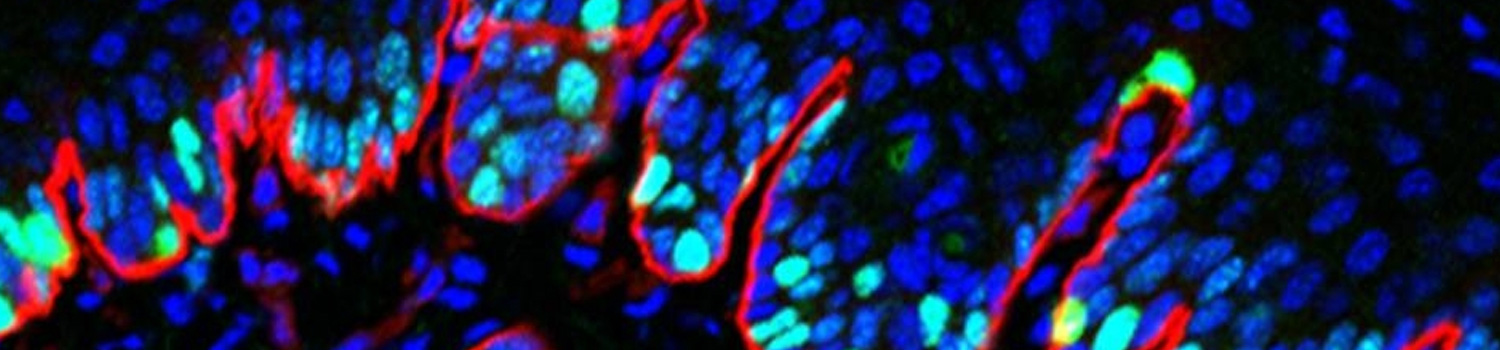
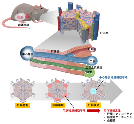
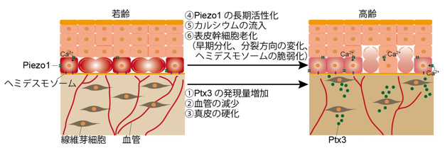
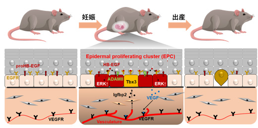
Crosstalk of various cells within each organ and networks between multiple tissues play important roles in maintaining homeostasis in our bodies. We are investigating the mechanisms of organ and tissue remodeling and their physiological functions to adapt to physiological changes that occur during life events and stages. We aim to understand the biological responses to maintain homeostasis at single-cell level, and applying the understanding to medical therapies.

Recent Achievement

Periportal hepatocyte proliferation at midgestation governs maternal glucose homeostasis in mice

The maternal liver is challenged by metabolic demands throughout pregnancy. However, hepatocyte dynamics and their physiological significance in pregnancy remain unclear. Here, we show in mice that hepatocyte proliferation is spatiotemporally regulated in each liver lobular zone during pregnancy, with transient proliferation of periportal and pericentral hepatocytes during mid and late gestation, respectively. Using adeno-associated virus (AAV)−8-mediated expression of the cell cycle inhibitor p21 in hepatocytes, we show that inhibition of hepatocyte proliferation during mid, but not late, gestation impairs liver growth. Transcriptionally, genes involved in glucose/glycogen metabolism are downregulated in late pregnancy when midgestational hepatocyte proliferation is attenuated. In addition, hepatic glycogen storage is abolished, with concomitant elevated blood glucose concentrations, glucose intolerance, placental glycogen deposition, and fetal overgrowth. Laser capture microdissection and RNA-seq analysis of each liver lobular zone show zone-specific changes in the transcriptome during pregnancy and identify genes that are periportally expressed at midgestation, including the hyaluronan-mediated motility receptor (Hmmr). Knockdown of Hmmr in hepatocytes by AAV8-shHmmr suppresses periportal hepatocyte proliferation at midgestation and induces impaired hepatic glycogen storage, glucose intolerance, placental glycogen deposition and fetal overgrowth. Our results suggest that periportal hepatocyte proliferation during midgestation is critical for maternal glycogen metabolism and fetal size.

Kozuki et al., Commun Biol 2023

Vasculature atrophy causes a stiffened microenvironment that augments epidermal stem cell differentiation in aged skin

Stem cell loss causes tissue deterioration associated with aging. The accumulation of genomic and oxidative stress-induced DNA damage is an intrinsic cue for stem cell loss; however, whether there is an external microenvironmental cue that triggers stem cell loss remains unclear. Here we report that the involution of skin vasculature causes dermal stiffening that augments the differentiation and hemidesmosome fragility of interfollicular epidermal stem cells (IFESCs) in aged mouse skin. Aging-related IFESC dysregulation occurs in plantar and tail skin, and is correlated with prolonged calcium influx, which is contributed by the mechanoresponsive ion channel Piezo1. Epidermal deletion of Piezo1 ameliorated IFESC dysregulation in aged skin, whereas Piezo1 activation augmented IFESC differentiation and hemidesmosome fragility in young mice. The dermis stiffened with age, which was accompanied by dermal vasculature atrophy. Conversely, induction of the dermal vasculature softened the dermis and ameliorated IFESC dysregulation in aged skin. Single-cell RNA sequencing of dermal fibroblasts identified an aging-associated anti-angiogenetic secretory molecule, pentraxin 3, which caused dermal sclerotization and IFESC dysregulation in aged skin. Our findings show that the vasculature softens the microenvironment for stem cell maintenance and provide a potential mechanobiology-based therapeutic strategy against skin disorders in aging.

Ichijo et al., Nat Aging 2022

Vasculature-driven stem cell population coordinates tissue scaling in dynamic organs

Stem cell (SC) proliferation and differentiation organize tissue homeostasis. However, how SCs regulate coordinate tissue scaling in dynamic organs remain unknown. Here, we delineate SC regulations in dynamic skin. We found that interfollicular epidermal SCs (IFESCs) shape basal epidermal proliferating clusters (EPCs) in expanding abdominal epidermis of pregnant mice and proliferating plantar epidermis. EPCs consist of IFESC-derived Tbx3+–basal cells (Tbx3+-BCs) and their neighboring cells where Adam8–extracellular signal–regulated kinase signaling is activated. Clonal lineage tracing revealed that Tbx3+-BC clones emerge in the abdominal epidermis during pregnancy, followed by differentiation after parturition. In the plantar epidermis, Tbx3+-BCs are sustained as long-lived SCs to maintain EPCs invariably. We showed that Tbx3+-BCs are vasculature-dependent IFESCs and identified mechanical stretch as an external cue for the vasculature-driven EPC formation. Our results uncover vasculature-mediated IFESC regulations, which explain how the epidermis adjusts its size in orchestration with dermal constituents in dynamic skin.

Ichijo et al., Sci Adv 2021

PAGE TOP